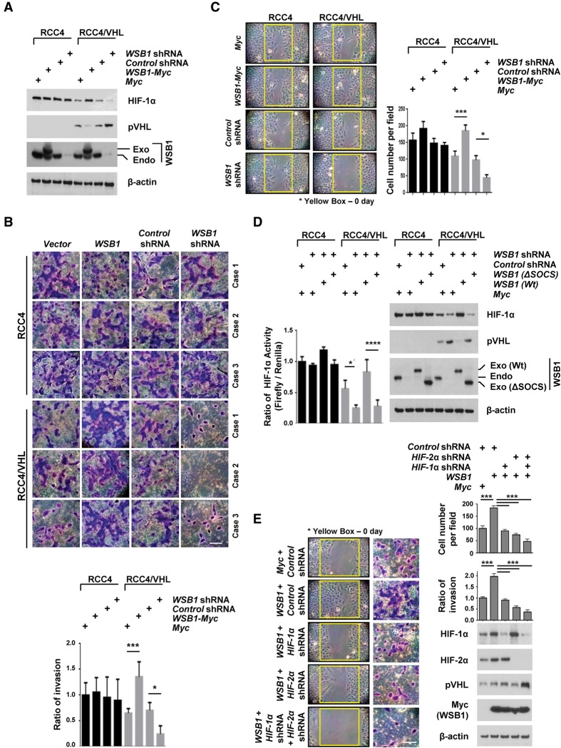
Figure 6.
WSB1 promotes cancer cell invasion and migration by inhibiting pVHL. (A) Cells were transfected with the indicated constructs, and pVHL and HIF-1α levels were examined. (Exo) Exogenous; (Endo) endogenous. (B) Trans-well invasion assay of RCC4 or RCC4/VHL cells stably transfected the indicated shRNAs or plasmids. The plot shows the quantification of the area covered by the invasion cells, relative to the control. The results represent the means ± SE of three independent experiments performed in triplicate. Bar, 10 μm. (*) P < 0.05; (***) P < 0.001 versus control cells by one-way ANOVA. (C) Wound healing assays of RCC4 or RCC4/VHL cell lines stably transfected with the indicated plasmids or shRNA. (Left) Representative images of the assay. (Right) The mean and SD of three independent experiments performed in triplicate are shown. (*) P < 0.05; (***) P < 0.001 versus control cells by one-way ANOVA. (D) RCC4 or RCC4/VHL cells were infected with the indicated constructs, and HIF-1α activity was assayed by a HIF-1α luciferase reporter assay. (Left) The results represent the means ± SE of three independent experiments performed in triplicate. (*) P < 0.05; (****) P < 0.0001 versus control cells by one-way ANOVA. (Right) Cells were blotted with the indicated antibodies. (Exo) Exogenous; (Endo) endogenous. (E) Cells were infected with indicated constructs. Wound healing assays and trans-well invasion assays of HIF-1α, HIF-2α, and pVHL levels were examined. (Left) Representative images of the wound healing assay. (Top right) The mean and SD of three independent experiments performed in triplicate are shown. (Middle) Representative images of the invasion assay. Bar, 10 μm. (Middle right) The mean and SD of three independent experiments performed in triplicate are shown. (Bottom right) Immunoblot with indicated antibodies. (***) P < 0.001 by one-way ANOVA.

