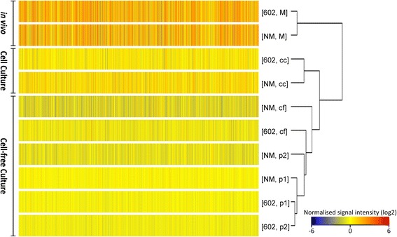
Fig. 2.

Hierarchical clustering analysis on normalised signal intensities of probes of 602 and NM strains in different culture models. Data shows clustering of in vivo and in vitro models separately based on patterns of gene expression. Normalised signal intensity (log2) of probes (average of triplicates) for each condition are represented as a colour scale from red for high expressions to blue for lower expressions. In vivo (M = mice spleen), cell culture (cc), cell-free adapting phase passage 1 (p1) and passage 2 (p2), cell-free culture model (cf)
