
Fig. 4.
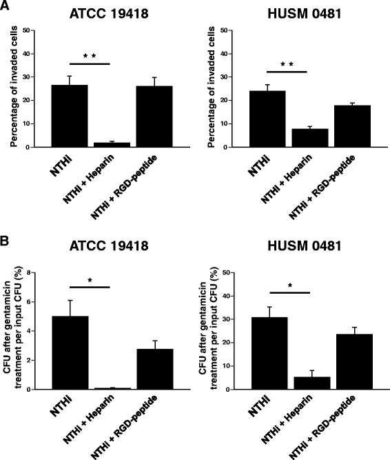
Intracellular invasion of NTHi in the presence of heparin or RGD peptide. BEAS-2B cells were infected for 2 hours with one of the two NTHi strains (ATCC 19418 or HUSM 0481), E. coli (a negative control), or L. monocytogenes (a positive control). In some experiments, cells were pretreated with heparin or RGD-peptide. a After killing extracellular bacteria with gentamicin, epithelial cells and bacteria were stained with LIVE/DEAD®. The percentages of BEAS-2B cells invaded by each type of bacteria are shown. b After killing extracellular bacteria with gentamicin and lysing the BEAS-2B cells, the bacteria were cultured overnight. The number of colonies was counted and the percentages of CFU after gentamicin treatment of cells per input CFU were shown. Error bars represent SEM in three independent experiments that gave similar results. *p < 0.05 and **p < 0.01