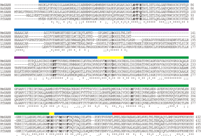
Figure 1. Amino acid sequence alignment of SAHHs.
Residues involved in nucleoside binding in MmSAHH are highlighted. The coloured lines above the sequence alignment represent the domains in MmSAHH. The domains are coloured for the catalytic (blue), coenzyme-binding (green), hinge (yellow), and C-terminal (red) domains. Insertion segments of 40 amino acid residues exist in MtSAHH, LlSAHH, and PfSAHH but not in mammalian SAHHs are indicated by a purple line. The abbreviations used are as follows: MmSAHH, Mus musculus SAHH; HsSAHH, Homo sapiens SAHH; MtSAHH, Mycobacterium tuberculosis SAHH; LlSAHH, Lupinus leteus SAHH; and PfSAHH, Plasmodium falciparum SAHH.

