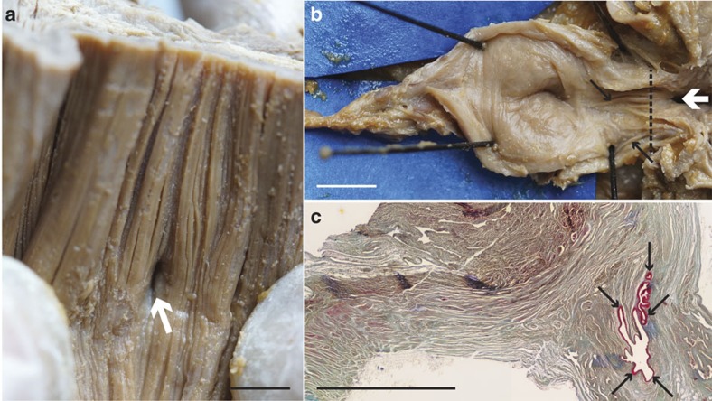
Figure 2. Evidence of pulmonary complex in L. chalumnae.
(a) Internal view of the ventral wall of the oesophagus showing the non-obliterated opening between oesophagus and lung in L. chalumnae CCC 3. Dorsal view of the oesophagus (anterior to the top). (b) Anterior part of the vestigial lung lumen from the dissection of the adult specimen CCC 3, ventral view, anterior to the right. (c) Histological thin section of the vestigial lung of CCC 5 in the region of the black dashed line in b. White arrows indicate the non-obliterated opening in specimen CCC 3, and black arrows indicate the invaginations of the internal wall of L. chalumnae vestigial lung in specimens CCC 3 and CCC 5. Scale bar, 0.5 cm (a–c).

