Figure 7.
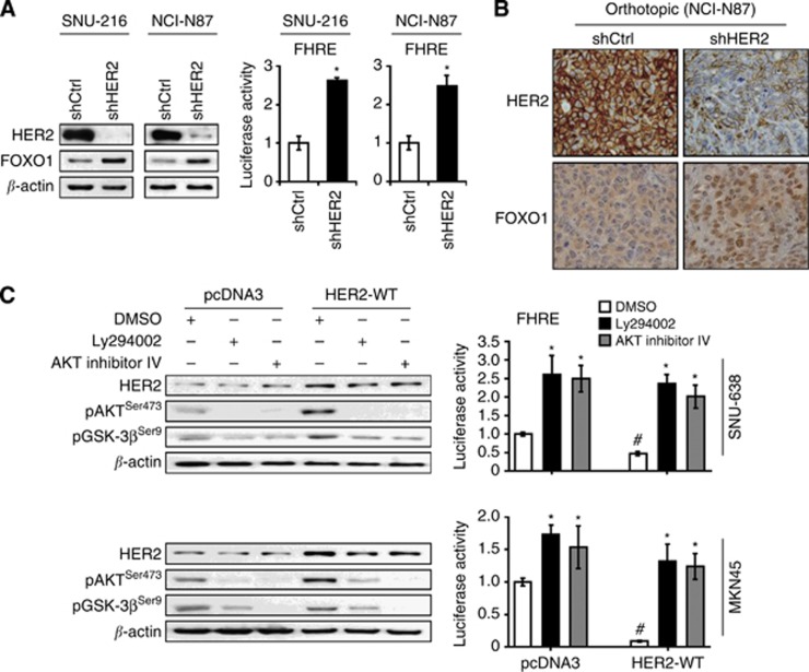
Effects of HER2 modulation on FOXO1 activation through PI3K/AKT pathway in GC cells and xenograft tumours. (A) SNU-216 and NCI-N87 cells were incubated with lentiviral particles containing a control shRNA (shCtrl) or HER2 shRNA (shHER2). The effects of HER2 silencing on FOXO1 expression and activation were determined by immunoblot analysis (left) and luciferase reporter assay (right), respectively. Luciferase activity in shCtrl cells was arbitrarily set to 1. Bars represent mean±s.d. (n=4). *P<0.05, compared with shCtrl. (B) Representative features of immunohistochemical staining for HER2 (upper panels) and FOXO1 (lower panels). Sections obtained from orthotopic xenograft tumours showed that shHER2-expressing tumours had higher FOXO1 expression in the nuclei of cancer cells than shCtrl-expressing tumours ( × 400 magnification). (C) SNU-638 and MKN45 cells were transfected with either a control plasmid pcDNA3 (pcDNA3) or an expression plasmid containing HER2 wild-type gene (HER2-WT). Cells were treated with a PI3K inhibitor LY294002 (30 μM) or an AKT inhibitor IV (0.5 μM) for 24 h, and immunoblot analysis was performed using the antibodies indicated in the figure (left). Luciferase reporter assay for FOXO1 transcriptional activity was also performed (right). Bars represent mean±s.d. (n=4). *P<0.05, compared with vehicle control (DMSO). #P<0.05, compared with vector control (pcDNA3).

