
Figure 3.
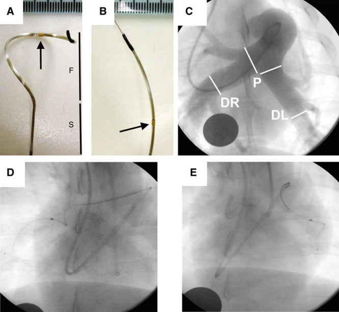
Pulmonary artery denervation (PDN) catheter and procedure. Prototype radiofrequency catheter (Medtronic Inc) off (A) and on (B) a 0.014″ guidewire (F indicates nitinol Spyral; S, catheter shaft; arrow, radiofrequency electrode; scale increment 1 mm). C, Pulmonary artery angiogram. White lines represent the proximal and distal boundaries for PDN procedure. Proximal (P) indicates pulmonary artery bifurcation; left distal (DL), ostium of the posterior artery; and right distal (DR), posterior descending artery (disc: 26 mm). Fluoroscopy of the PDN catheter in the right (D) and left pulmonary artery (E).