Figure 2.
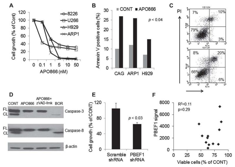
APO866 and PBEF1 knockdown induces apoptosis and inhibits growth of myeloma cells, (A) The indicated MM cell lines were treated with increasing concentrations of APO866 for 72 hours and then subjected to MTT assay. (B, C) The indicated MM cell lines were treated with APO866 (2.5 nmol/L, 48 hours) and then subjected to apoptosis assay with annex in V–PI flow cytometry. Percentages of annex in V–positive (apoptotic) cells (B) and a representative flow cytometry analysis of ARP1 cells (C) are shown. (D) Western blot analysis demonstrating lack of cleavage (activity) of caspase-3 or caspase-8 in the absence or presence of z-VAD-fmk (pan caspase inhibitor) in ARP1 cells exposed to APO866 for 24 hours. The proteasome inhibitor bortezomib (BOR) was used as a positive control measure. (E) Luciferase-expressing ARP1 MM cells were infected with lentiviral particles containing scramble or PBEF1 shRNA and then subjected to growth assay using luciferase activity (96 hours). (F) Freshly obtained CD138-selected MM cells from 13 patients were treated with APO866 (10 nmol/L) for 96 hours. The effect on MM cell survival and growth was assessed by trypan blue exclusion, whereas PBEF1 gene expression was determined by GEP. Overall, APO866 inhibited survival and growth of primary MM cells by 49% ± 4% (p < 0.0001), an effect that was not correlated with expression of PBEF1 in these cells.

