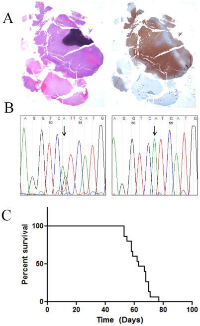
Fig. 1. IDH1 mutation, mutant protein expression and in vivo growth JHH-273.
(A) H&E staining of resected patient tumor tissue showed areas of hypercellularity and mitotic figures (left) as well as strong IDH1 (R132H) protein expression (right) leading to a diagnosis of IDH1 mutant anaplastic astrocytoma (WHO grade III) (B) Sequencing of exon 4 of IDH1 shows the original heterozygous G395A (R132H) mutation (left) in the original patient tumor, with loss of the wild type copy in the first passage xenograft (right) (C) Orthotopically implanted xenografts have a median survival time of 63 days and are uniformly lethal (n=15).

