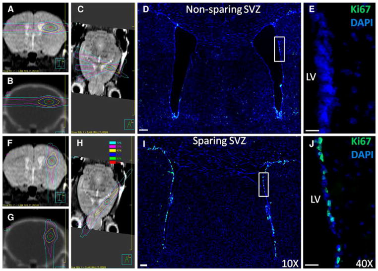
Fig. 1.
Mouse radiation treatment plans (left) and microscopy images (right) for the non-NPC-sparing (top) and NPC-sparing (bottom) radiotherapy plans. Left side MRI and CT images from the mouse radiation treatment plans showing the radiation dose distribution for the non-NPC sparing (top; a–c) and NPC-sparing radiation treatment plans (bottom; f–h). Note that for the non-NPC sparing plan, the region of the SVZ of the ipsilateral lateral ventricle receives a high radiation dose, whereas this region is effectively spared in the NPC Scans are: coronal MRI (a, f), coronal CT (b, g), and axial MRI (c, h). Dose values are shown in the legend. Right side Coronal sections showing Ki-67 stains (green) in the SVZ of the lateral ventricles following non-NPC sparing RT (d, e) and NPC-sparing RT (i, j). Co-staining is with DAPI (blue). Images d and i were taken with a 10× objective and the images e and j with 40× objective. LV left ventricle. (Color figure online)

