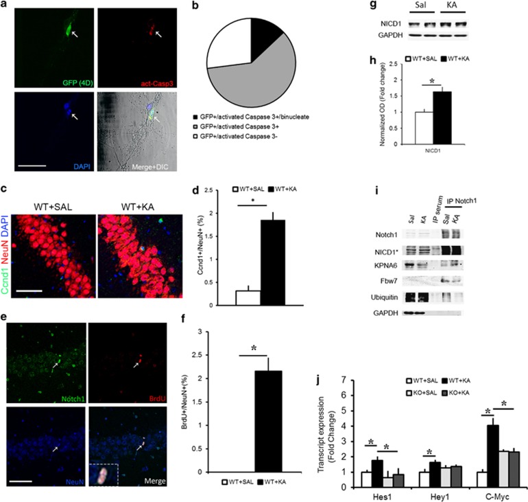
Figure 1.
Cell cycle reentry following KA insult is associated with nuclear Notch signaling. (a) Representative fluorescence immunohistochemistry photomicrographs of primary neuronal cultures transfected with a Ccnd1:CDK4 (4D) construct colabeled with act-Casp3 and DAPI. Arrow indicates a binucleate 4D+ neuron. (b) Pie chart summarizing the proportion of neurons labeled with GFP only, those colabeled with act-Casp3 or those colabeled with act-Casp3 and with binucleate morphology (n=50 cells). (c) Representative immunofluorescence photomicrograph of Ccnd1 expressing neuron from the WT hippocampi treated with KA. (d) Quantitation of the percentage of Ccnd1-positive neurons in Saline and KA hippocampi (n=4 WT mice per condition). (e) Hippocampal CA1 layer, 2 days following KA insult, shows the presence of BrdU+ neurons expressing Notch1 in the nucleus (white arrow and insert). (f) Graph summarizing the proportion of BrdU+ cells as a percentage of NeuN+ neurons in the CA layer following KA as compared with Saline controls (n=951 cells from n=4 mice per condition). (g) Representative immunoblot shows the extent of cleaved Notch1 in hippocampal lysates at 12 h after KA as compared with Saline controls. (h) Optical density quantitation of the NICD1 bands 12 h after Saline and KA injection (n=8 per condition). (i) Representative immunoblot on immunoprecipitated hippocampal lysates using an antibody against the cytoplasmic tail of Notch1 shows the following interactions- Notch1:Kpna6 and Notch1:fbw7, and ubiquitination of Notch1 in Saline and KA conditions. No trace of protein is visible in the control IP sample with serum. GAPDH is used to control for loading of the inputs and contamination in the IP samples (n=4 independent IPs). (j) Graph summarizing the expression of the Notch targets; Hes1, Hey1 and C-myc, upon KA in WT and RBPJKcKO hippocampi as compared with the respective Saline controls (n=4-8 mice per condition). Asterisks indicate significant differences. Error bars represent mean±S.E.M. Scale bars in A and C is 50 μm and E is 100 μm

