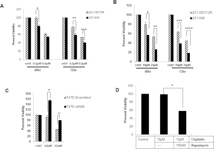
Figure 3. NMI expression increases chemosensitivity.
(A) MDA-MB-231 vector or NMI cells were plated in a 96 well plate in triplicate and treated with either 0.1 μM or 0.5 μM doxorubicin for 48 and 72 hours and MTS was done to measure cell viability. Percent viability is measured as fold change of corresponding vehicle control (*p = .04, **p = .04, ***p = .02) (B) Cisplatin treatment of either 10 μM or 25 μM at 48 and 72 hours (*p = .03, **p = .003, ***p = .002, ****p = .0001) (C) T47D scrambled or shNMI cells were treated with 10 μM or 25 μM cisplatin for 48 hours and MTS was done to measure cell viability. (*p = .01, **p = .005). (D) T47D NMI silenced cells were treated with 10 μM cisplatin alone or in combination with 100 nM rapamycin for 48 hours and MTS was done to measure cell viability (*p = .001).

