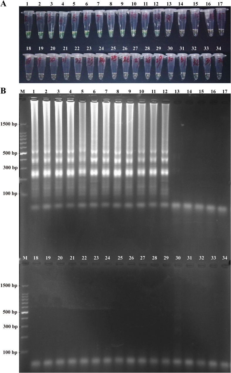
Figure 9. Specificity of MCDA detection for different strains.
(A) The MCDA reactions were visualized as color change by FD reagent. (B) The MCDA products were observed as ladder-like pattern by 2.5% agarose gel electrophoresis. Tube (Lane) 1–12, L. monocytogenes strains of serovar 1/2a (EGD-e), 1/2b (ICDCLM007), 1/2c (ICDCLM010), 3a (ICDCLM023), 3b (ICDCLM078), 3c (ICDCLM446), 7 (NCTC10890), 4a (ATCC19114), 4b (ICDC419), 4c (ATCC19116), 4d (ATCC19117) and 4e (ATCC19118); tube (lane) 13-17, others Listeria reference strains of L. innocua (ATCCBAA-680), L. ivanovii (ATCCBAA-678), L. seeligeri (ATCC35967), L. welshimeri (ATCC35897), L. grayi (ATCC25402); tube (lane) 18-34, non-Listeria strains of Enteropathogenic E. coli, Enterotoxigenic E. coli, Enteroaggregative E. coli, Enteroinvasive E. coli, Enterohemorrhagic E. coli, Enterobacter cloacae, Enterococcus faecalis, Bacillus cereus, Vibrio vulnificus, Vibrio fluvialis, Vibrio parahaemolyticus, Yersinia enterocolitica, Streptococcus pneumonia, Shigella flexneri, Plesiomonas shigelloides, Salmonella enteric, Klebsiella pneumonia. Lane M, DL 100-bp DNA marker.

