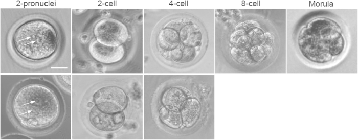
Figure 3. Embryonic development of two-pronuclei embryos obtained by parthenogenetic activation (PA, top row) and in vitro fertilization (IVF, bottom row) cultured on the feeder layer of inactivated mouse embryonic fibroblasts (MEFs) in embryonic stem cell (ESC) medium, showing the development to 4-cell stage of the parthenogenetically activated and in vitro fertilized embryos, respectively.
The parthenogenetically activated oocytes could further develop to 8-cell and morula stages. Arrows indicate pronuclei. Scale bar: 15 μm.

