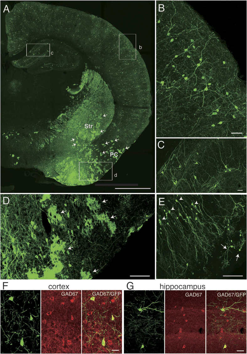
Figure 1. IUE labeled only neurons in the neocortex, hippocampus and olfactory bulb but also labeled astrocyte-like cells in the subpallium.
A, Coronal section at the level that includes the striatum and hippocampus. Arrows point to cloud-like structures in the striatum (Str) and the piriform cortex (PC), which were identified as astrocytes using high-resolution views (see Fig. 2). B, High-magnification view of the area shown by the rectangle labeled “b” in A. Cells with neuronal morphology are labeled in the neocortex. C, High-magnification view of the area shown by the rectangle labeled as “c” in A, showing labeled cells with neuronal morphology in the hippocampus. Note the absence of cloud-like profiles either in the neocortex or the hippocampus. D, High-magnification view of the area shown by the rectangle “d” in A. Many cloud-like cells that appear to be astrocytes are labeled in the piriform cortex (arrows). E, Granule cells (arrowheads) and periglomerular cells (arrows) in the olfactory bulb. No astrocyte-like cells can be seen. F, G, GABAergic nature of labeled neurons in the neocortex (F) and hippocampus (G). Left panels in F and G show GFP-labeled cells, middle panels GAD67 immunolabeled cells and right panels merged views. Scale bars: 1 mm in A, 40 μm in B and C, 100 μm in D and E, and 20 μm in F and G.

