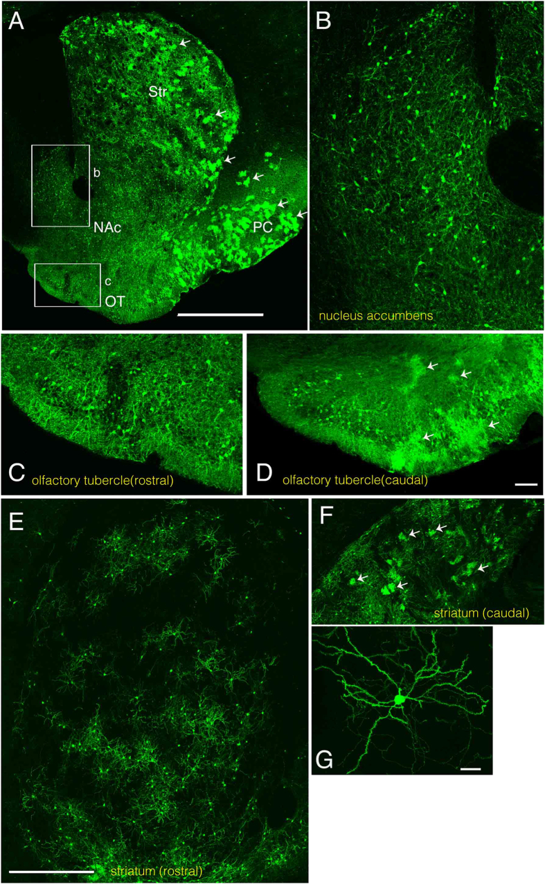
Figure 3. IUE-labeled neurons and astrocytes in the nucleus accumbens, olfactory tubercle, and striatum.
A, Low-magnification image showing labeled striatum (Str), nucleus accumbens (NAc), olfactory tubercle (OT) and piriform cortex (PC). Many astrocytes are labeled in the striatum and the piriform cortex (arrows). Lateral is to the right and ventral is to the bottom. B,C, High magnification views of the boxed areas labeled “b” and “c” in A show regions of the nucleus accumbens shell region and olfactory tubercle, respectively. D, Olfactory tubercle at a caudal level of the same animal. While no cloud-like profiles can be detected at the rostral level (C), there are many at the caudal level (D), indicating an abundance of astrocytes there. In some embryos, the rostral striatum was almost exclusively occupied by neurons (E). In the caudal striatum, both neurons and astrocytes are also labeled (F)(arrows). G, Most labeled neurons in the striatum are medium spiny neurons. In E-G, lateral is to the left and ventral is to the bottom. Scale bars: 1 mm in A, 100 μm in D, 500 μm in E and 20 μm in G. The bar in E also applies to F.

