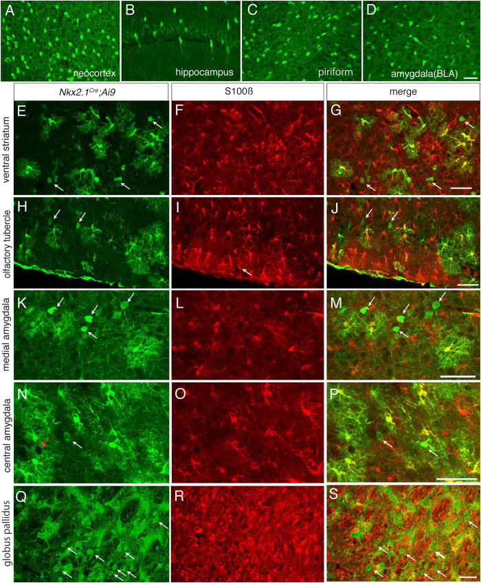
Figure 5. Labeled neurons and astrocytes in Nkx2.1Cre; Ai9 mice.
A–D, Labeled cells in the neocortex (A), hippocampus (B), piriform cortex (C) and basolateral amygdala (D). None of the labeled profiles have ramified processes, suggesting that they are not astrocytes. E–S, Neurons and astrocytes labeled in the ventral striatum (E,G), olfactory tubercle (H,J), medial amygdala (K,M), central amygdala (N,P) and globus pallidus (Q,S). Unlike A–D, cells with ramified processes are intermingled with neuron-like cells (arrows in E,H,K,N,Q,G,J,M,P and S). The former are positive for S100 beta (F,I,L,O,R), an astrocyte marker. G,J,M,P and S are merged views showing double labeled cells in yellow. Red arrow in N points to a hole-like structure. In A,B,C,D,E,H,K,N and Q, red signal of tdTomato was converted to green. Scale bar: 50 μm for all panels. The bar in D applies to A–D, that in G to E–G, that in J to H–J, that in M to K–M, that in P to N–P and that in S to Q–S.

