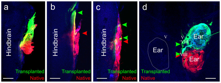
Figure 3. Inner ear afferent projections.
(a) Animal in which inner ear afferents projected entirely together in the VIIIth ganglion. (b) Animal in which inner ear afferents from the natively-oriented transplanted ear projected in their own ‘VIIIth’ ganglion and entered the hindbrain separate from the inner ear afferents from the native VIIIth ganglion. (c) Animal in which the inner ear afferents from the 90 degrees rotated transplanted ear entered the hindbrain from both in its own ‘VIIIth’ ganglion and with the native VIIIth ganglion. (d) Animal in which the inner ear afferents leave the natively-oriented transplanted ear both in its own ‘VIIIth’ ganglion and along with the native VIIIth ganglion. Green arrowheads indicate projections from the transplanted ear. Red arrowheads indicate projections from the native ear. V, trigeminal nerve. Scale bar is 100 µm.

