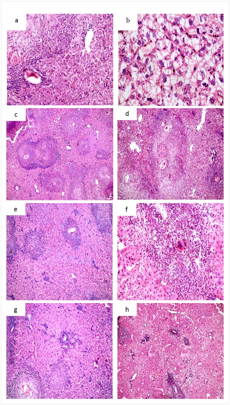
Fig 5. Histopathological study of H&E stained liver sections of different groups of mice infected with Schistosoma mansoni.
Infected untreated mice showing:(a) preserved hepatic acinar architecture, intense inflammatory infiltration and Kupffer cell hyperplasia (X200); (b) Marked ballooning and swelling of hepatocytes (X400); (c), (d) and (e) several granulomas consisting of epithelioid, eosinophilic and lymphocytic cells surrounding well developed eggs (X100, X200 and 400 respectively). Infected, MFS-LNCS-OA-treated mice showing: (f), (g) and (h) small cellular granulomas with mild degree of cellular swelling and inflammatory infiltration (X100, X200 and X100 respectively).

