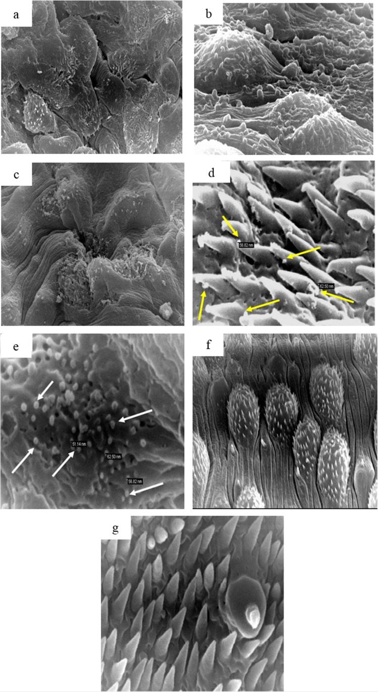
Fig 6. Scanning Electron Microscopy (SEM) of a male Schistosoma mansoni worm from a mouse treated with MFS-LNC-OA showing.
(a) Marked tegumental irregularity and disfigurement (X 3,500); (b) Tegumental surface blebbing (X 7,500); (c) Edema, flattening and sloughing of the whole tubercles with partial to complete loss of the spines (X 5,000); (d) and (e) nano-objects of similar size to lipid nanocapsules in between spines and on damaged schistosomal surface, respectively (X35,000). SEM of a normal male worm showing: (f) and (g) normal dorsal tegumental surface and papilla (X5,000 and 35,000 respectively)

