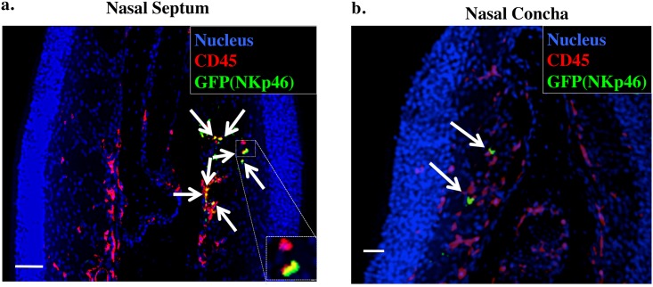
Fig 1. Visualization of nasal NKp46+ cells by using immunohistochemistry.
Frozen sections of nasal tissue obtained from 8-week-old Ncr1 GFP/+ mouse were stained with 4′,6-diamidino-2-phenylindole (nucleus), anti-CD45, and anti-GFP antibodies and examined under a fluorescence microscope. Arrows indicate CD45+GFP(NKp46)+ cells. Bar, 50 μm. Data are representative of at least 3 independent experiments. a. Nasal septum. b. Nasal concha.

