Figure 4.
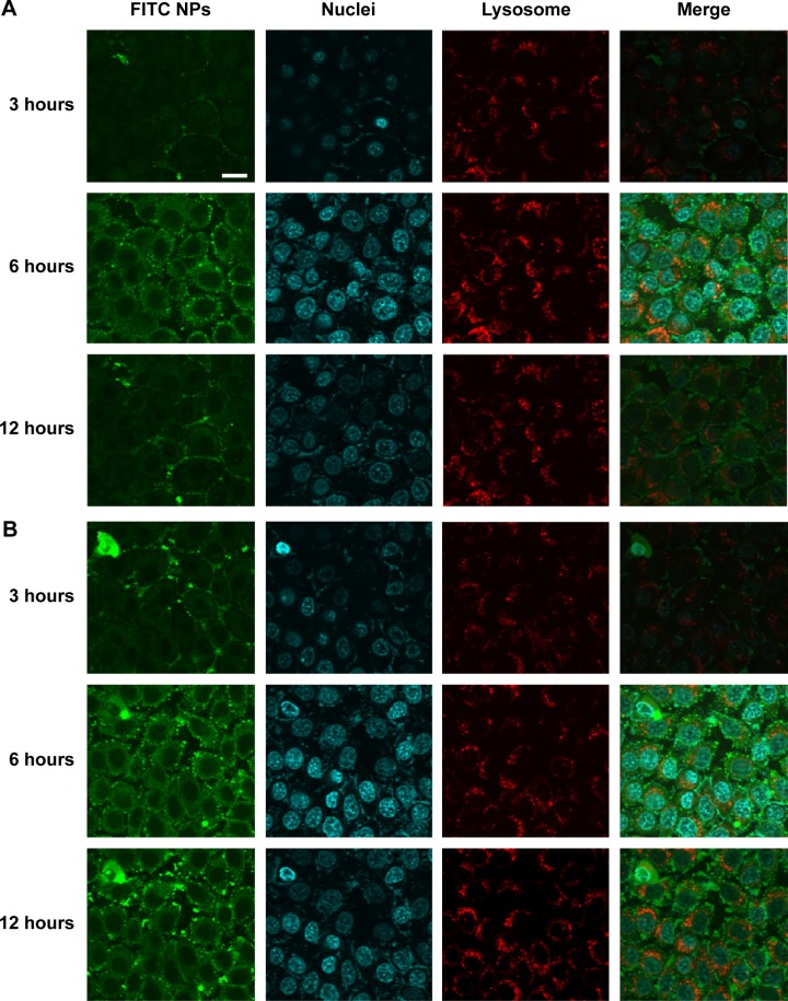
Confocal images of (A) FITC labeled blank NPs and (B) FITC labeled shMDR1 NPs incubated with cells for 12 hours.
Notes: The nucleus was stained with Hoechst (blue) for 15 minutes at 37°C and all NPs were labeled with FITC (green). The lysosome was stained with LysoTracker Red DND-99. The scale bar is 50 μm and applies to all figure parts.
Abbreviation: FITC, fluorescein isothiocyanate.

