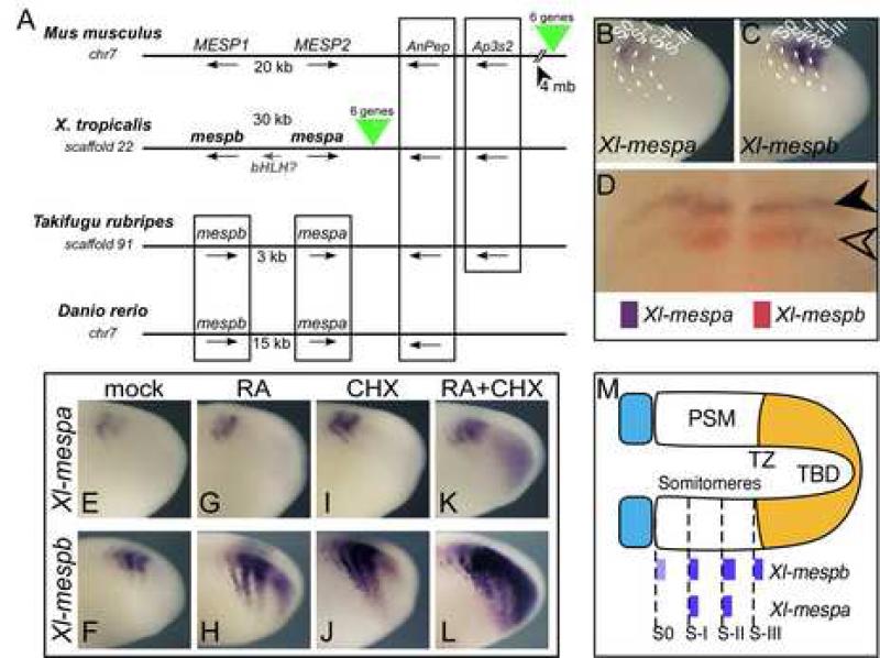
Figure 1.
Mesp genes in Xenopus. (A) Genomic organization of the Mesp genes. Shown are syntenic genomic regions from mouse, Xenopus tropicalis, Takifugu rubripes, and Danio rerio. Chromosome number is given when available, otherwise, scaffold number is indicated. Boxing indicates synteny of genes. X. tropicalis genes ENSXETG00000017725 and ENSXETG00000027628 are designated as mespb and mespa respectively. Green triangles indicate a syntenic block of 6 genes that are conserved between X. tropicalis and mouse, but in frog this block is between mespa and the AnPep gene, while in mouse the block is located 4 megabases (mb) away. (B-C) Expression of Xl-mespa and Xl-mespb in the PSM of X. laevis embryos by whole mount in situ hybridization. Dotted lines indicate approximate locations of somitomere boundaries. (D) Double in situ hybridization with Xl-mespb (red) and Xl-mespa (purple). (E-L) Shown is the expression of Xenopus Mesp genes after treatment with CHX and RA, as indicated. Treatment was for 1.5 h at RT prior to fixation. (M) Schematic representation of the PSM showing the relative locations of the somitomeres, transition zone (TZ), and tailbud (TBD) with Xenopus Mesp expression patterns indicated. Anterior is to the left in all panels.

