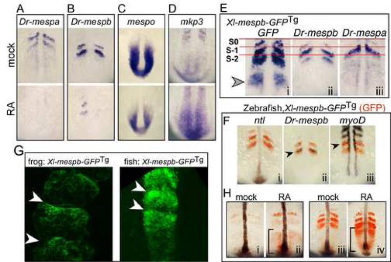
Figure 5.
Zebrafish Mesp genes respond differently to RA treatment. (A-D) Zebrafish embryos treated with carrier control (DMSO; top panels) or with RA (lower panels) for 3 hours and then stained for the expression of the indicated gene: A, Dr-mespa; B, Dr-mespb; C, mespo; D, mkp3. (E) Zebrafish embryos transgenic for Xl-mespb-GFP were stained for GFP RNA (i), Dr-mespb RNA (ii) or Dr-mespa RNA expression (iii) an aligned according to the first somite. (F) Double-label in situs on zebrafish embryos transgenic for XMespβ–GFP with GFP (red) and the gene indicated (purple) to localize the GFP expression pattern. (G). X. laevis (left panel) and zebrafish embryos (right panel) transgenic for the full-length Xl-mespb-GFP were imaged by confocal microscopy. Shown is a region containing newly formed somites, and where somitic boundaries are marked with arrowheads (H) Zebrafish embryos transgenic for Xl-mespb-GFP were treated with RA at the 6 somite stage (i,ii) or 12 somite stage (iii, iv) and stained for GFP RNA (red) and for no tail (ntl) in purple to mark the notochord. Brackets mark the tailbud domain. Anterior is oriented up in all panels.

