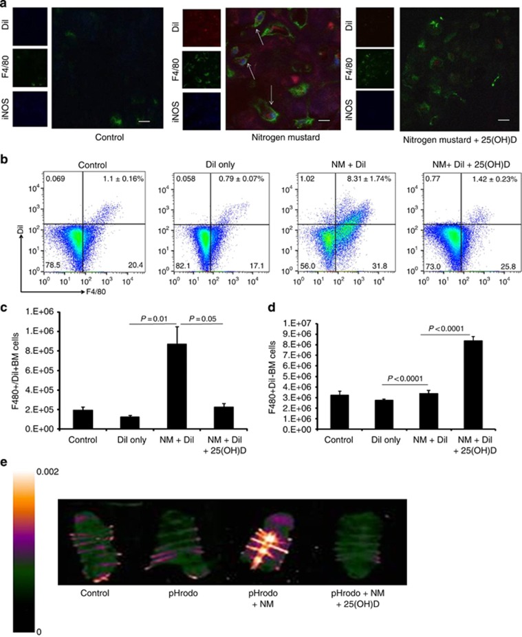
Figure 4.
Appearance of dermal hyperactivated macrophages in the BM after topical NM exposure. NM-exposed mice were injected intradermally with DiI post NM exposure with and without 25(OH)D. (a) Sternums were sectioned for co-localization of DiI (red), iNOS (blue) and F4/80+ (green) macrophages in the marrow (arrows) by confocal microscopy. Bone marrow cells were subjected to flow cytometric analysis (b) to detect F4/80+DiI+ and F480+DiI− macrophages, and for quantitative analysis of absolute cell numbers of (c) F4/80+ DiI+ cells (n=4; P=0.01, P=0.05) and (d) F4/80+DiI− cells (P<0.0001). pHrodo dye conjugated with bacteria was injected subcutaneously at the wound site 1 hour following NM exposure to detect (e) pHrodo-derived fluorescence in the sternum of NM-exposed mice 5 days after injection as visualized by Maestro imaging. Scale Bar=100 μm.

