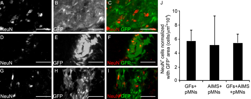
Figure 4. pMNs differentiate into neurons when transplanted into the injured spinal cord.
(A–I) Representative images from GFs+pMNs, GFs+AIMS+pMNs, and AIMS+pMNs groups stained with the neuronal nuclei marker, NeuN, two weeks post-transplantation. (A–C) GFs+pMNs group showing NeuN+ cells (A) and GFP+ areas (B) that colocalize together (C). (D–F) GFs+AIMS+pMNs groups also stained positive with NeuN (D) within GFP+ areas (E) and showed colocalization (F). (G–I) AIMS+pMNs also showed NeuN+ cells (G) within GFP+ areas (H) which colocalize together (I). (J) The GFs+pMNs group had significantly greater GFP+ area and higher numbers of NeuN+ cells, but when NeuN+ cell number is normalized by the GFP+ area similar levels of NeuN expression between all groups. Scale bars are 100 µm. Error bars are standard deviation.

