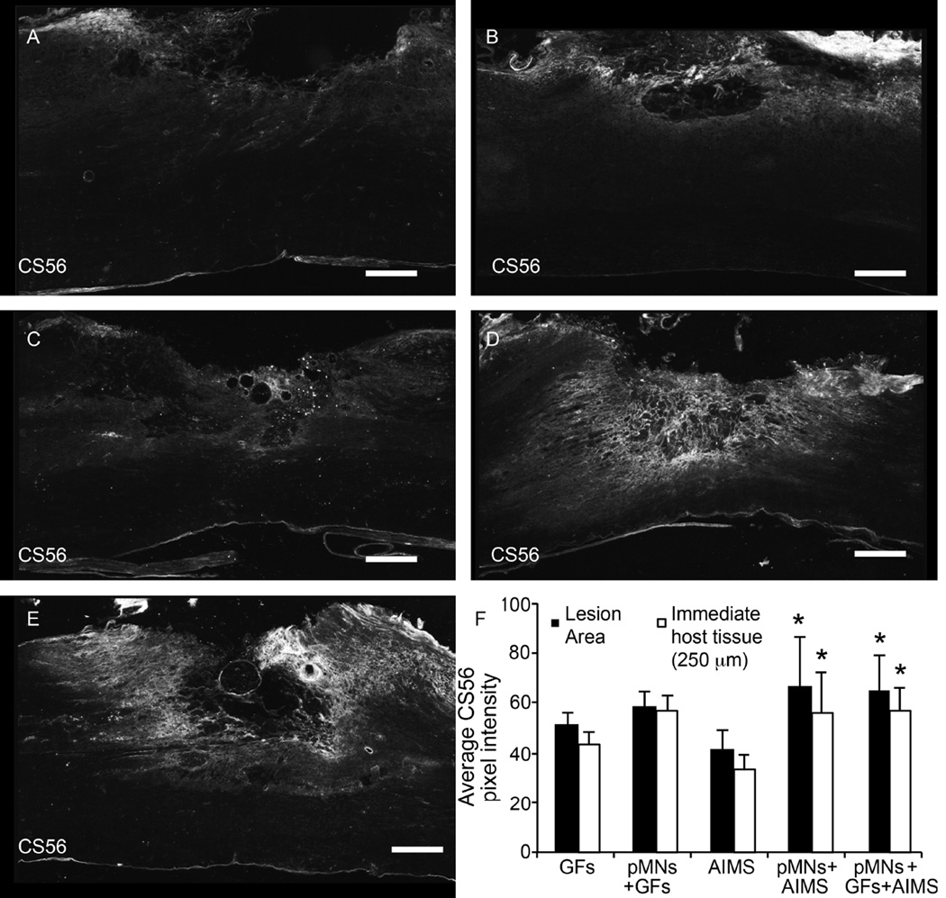
Figure 7. Combination of pMNs and/or GFs with AIMS does not decrease CSPGs compared to GFs alone or pMNs+GFs.
(A–E) Representative images of CS56, a marker for non-degraded CSPGs, staining within and around the lesion site for groups containing (A) GFs, (B) pMNs+GFs, (C) AIMS, (D) pMNs+AIMS, (E) pMNs+GFs+AIMS. Strong CS56 staining was seen when pMNs and AIMS were included in fibrin scaffolds. (F) The average pixel intensity of CS56 stained sections within the lesion area and within the immediate host tissue (250 µm from the lesion border) was determined. Fibrin scaffolds with pMNs+AIMS or pMNs+GFs+AIMS had significantly higher CS56 staining compared to the AIMS alone group in both the lesion area and immediate host tissue. Error bars are standard deviation. * denotes p<0.05 versus AIMS alone group.

