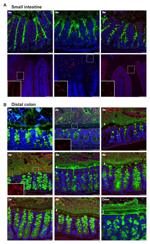
Figure 3. Immunohistology of small intestine and colon during conventionalization.
Immunostaining of sections from distal small intestine (A) and distal colon (B) of Muc2 (green) with FISH detecting bacteria (red) and Hoechst counterstain of DNA (blue). Double arrows indicate the inner mucus layer. Inserts show the bacteria magnified in the boxed areas. Typically, the middle part of the inner mucus layer is not stained as well as Muc2 at other locations. Scale bars in A, upper panel and B, 50 μm; A, lower panel, 25 μm.

