
Figure 2. Vemurafenib induces the release and secretion of TGF-β from some BRAF-mutant melanoma cells.
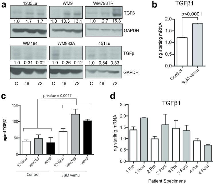
A Western blot analysis of 6 melanoma cell lines treated with vemurafenib (3 μM, 72Hrs). Densitometry for TGF-β is depicted in fold changes compared to each respective control. Scale bar = 50 μm. B: qRT-PCR for TGF-β1 shows vemurafenib-mediated induction of TGF-β1 mRNA expression in 1205Lu. Data was normalized to GAPDH and 18S endogenous controls. C: ELISA showing induction of TGF-β release from BRAFV600E melanoma cell lines following 3μM vemurafenib treatment (72 hours), expressed in pg/ml. D: Data shows q-RT-PCR experiments measuring levels of TGF-β1 mRNA in 4 matched (pre and post treatment) pairs of melanoma patient specimens receiving vemurafenib therapy (960 mg BID), error bars represent technical replicates of a single RNA extraction.