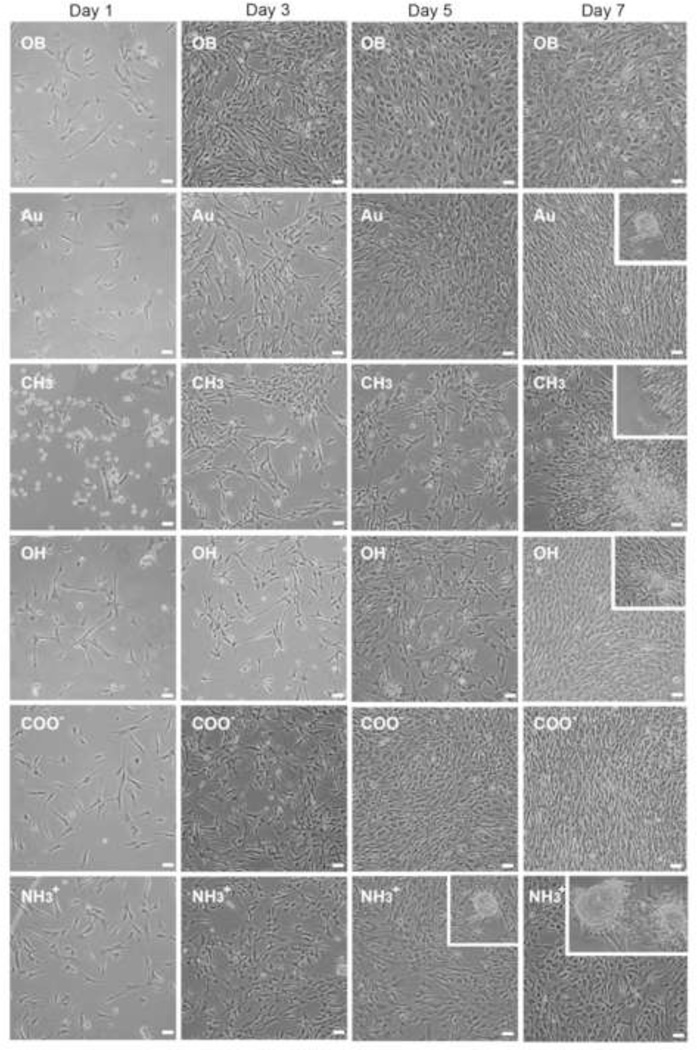
Figure 2.
Valvular interstitial cell (VIC) proliferation on monolayer surfaces as compared to bare glass/osteoblastic (OB) and gold controls (dashed lines). Initial inhibition of VIC growth (1–3 days) is exhibited by OH and CH3-SAMs with significant lower cell concentration, while osteoblastic (OB) controls are significantly greater than other treatments. Between 3 and 5 days, VICs on OH-SAMs begin proliferation while CH3-SAMs result in significantly lower cell concentration throughout the experiment. By day 5, COO−-SAMs have significantly higher cell concentrations than any other treatment. Between five and seven days in culture all samples, except CH3-SAMs, reach confluence. *p < 0.05, n = 6.

