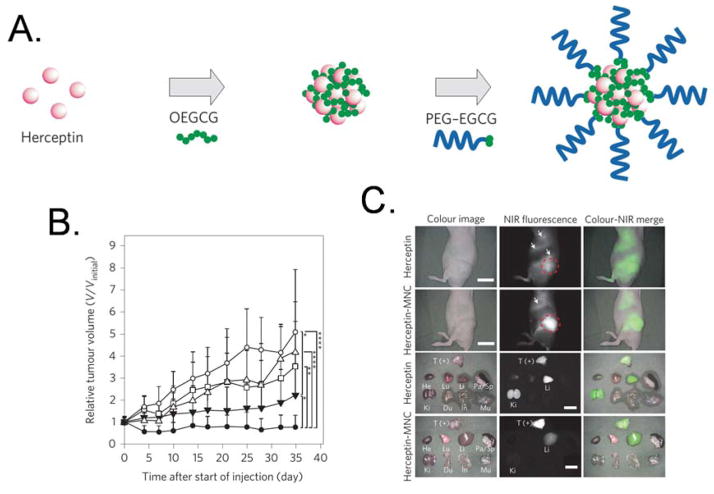
Figure 4.
a) Schematic diagram of the self-assembly process used to form the micellar nanocomplexes, which are formed via two sequential self-assemblies in an aqueous solution: complexation of OEGCG with proteins to form the core, followed by complexation of PEG–EGCG surrounding the preformed core to form the shell. b) Anticancer effect on BT-474-xenografted nude mouse model. PBS (vehicle control, open circles), BSA–MNC (open triangles), Herceptin (2.5 mg/kg, open squares), sequential injection of BSA–MNC and Herceptin (filled inverted triangles) and Herceptin–MNC (filled circles). c) Real-time intraoperative tumor detection and NIR fluorescence image-guided resection at 24 h post-injection. Arrows indicate nonspecific uptake (liver, kidneys, intestine). The red dashed circle delineates the region of interest. Abbreviations used are: BSA, bovine serum albumin; EGCG, Epigallocatechin-3-O-gallate; MNC, micellar nanocomplex; OEGCG, oligomerized EGCG; PEG, polyethylene glycol; T (+), positive tumor. Reprinted with permission from ref. 78. Copyright 2014 Nature Publishing Group.

