FIGURE 4.
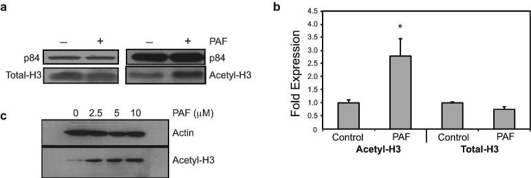
Effect of PAF on histone acetylation. (a) Protein levels of total-H3 and acetylated-H3 in HMC-1 cells 24 hours post cPAF (10 μM) treatment. p84 is the loading control. (b) Acetylation of CXCR4 promoter by ChIP analysis. HMC-1 cells were treated with 10 μM cPAF and harvested 24 hours later. Total-H3 was used as positive control and the data were normalized against input DNA. Data represent the mean ± SEM (N=4), *p < 0.05 vs. control (Mann-Whitney U-test,). (c) PAF up-regulates the expression of acetyl-H3 mRNA in normal mast cells. Buffy coat-derived mast cells were treated with increasing concentrations of cPAF and immunoblotting was used to determine the expression of acetyl-H3(K9/14/18/23/27). Actin is the loading control.

