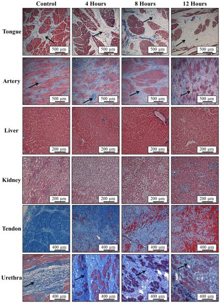
Figure 4. Histology: 58°C Samples.
Images show histology slides stained with a trichrome blue staining for samples heated at 58°C for 4, 8, and 12 hours. Results for tongue, artery, and tendon demonstrated a decrease in collagen (blue) density with heating. Furthermore, some hydrolysis into gelatin (red) was observed in tendon after 8 and 12 hours. Urethra results demonstrated an increase in collagen density after heating compared to control.

