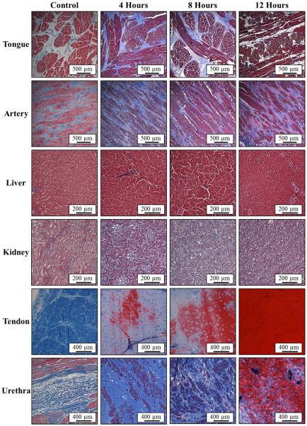
Figure 6. Histology: 90°C Samples.
Images show histology slides stained with a trichrome blue staining for samples heated at 90°C for 4, 8, and 12 hours. Results for tongue, artery, and urethra demonstrated an increase in collagen (blue) density with heating. In tendon, collagen was observed to hydrolyze into gelatin (red) with heating. Some hydrolysis was also observed in the urethra after 12 hour heating.

