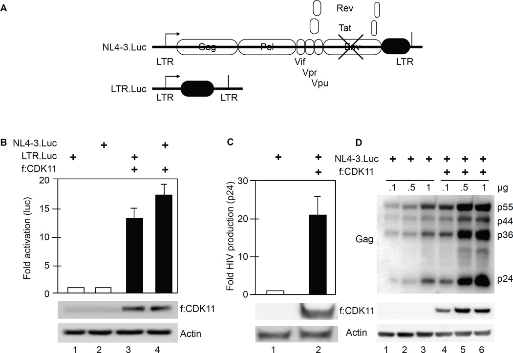
Figure 2. CDK11 increases HIV gene expression and replication in 293 T cells.
A. Schematic representation of NL4-3.Luc and LTR.Luc plasmid targets, respectively. NL4-3.Luc does not express Env (crossed lines) and contains the luciferase gene in the place of Nef.
B. CDK11 activates NL4-3.luc and LTR.Luc. Luciferase activity was measured in lysates of cells co-expressing f:CDK11 or the empty plasmid vector and LTR.Luc or pNL4-3.Luc. Error bars represent mean ± SE, n = 3. Western blots of f:CDK11 and actin as the loading control are presented below the bar graph. See also Figures S1 and S2.
C. CDK11 increases the production of new viral particles. Presented is the HIV p24 ELISA of supernatants from cells that co-expressed f:CDK11 and NL4-3.Luc. Error bars are as in panel B.
D. CDK11 increases the production of HIV structural proteins. Presented is a western blot of HIV Gag levels in cells expressing f:CDK11 and NL4-3.Luc or plasmid vector control. Amounts of plasmid effectors are presented above the western blot. Lanes 1 and 4, 2 and 5 and 3 and 6 should be compared. Below this panel are western blots of f:CDK11 and actin, as in panel C.

