Figure 1.
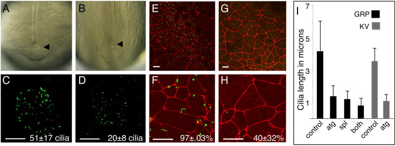
Knock down of FoxJ1 activity inhibits ciliogenesis in the Zebrafish KV and Xenopus GRP.
(A-B) KV (arrowhead) morphology was visualized in Zebrafish embryos using light field microscopy in control (A) ZFoxJ1 morphants (B). (C-D) Cilia in KV were visualized by staining with the acetylated tubulin antibody (green) and confocal microscopy in control (C) and ZFoxJ1 morphants (D). Average number of cilia per KV (n=12) is indicated (N±S.D.) (E-H) Dorsal explants were generated from stage 17 Xenopus embryos injected with a mixture of XFoxJ1-MOATG and XFoxJ1-MOSPL (G,H) or with a control morpholino (E,F) and stained with ZO-1 (red) and acetylated tubulin (green) antibodies, to label cell junctions and cilia, respectively. The percentage of GRP cells (n=100-120 cells from 6 embryos) that extend cilia is indicated (%ciliated ±S.D.) Scale bars=20μm in all panels. (I) Cilia length (n=100-120 cilia from 6 embryos) was measured on the GRP of Xenopus embryos injected with a control-MO (control), with XFoxJ1-MOatg (atg), with XFoxJ1-MOSPL (spl), or with a mixture of both XFoxJ1-MOs (both). Cilia length in the KV of Zebrafish embryos (n=20 cilia from each of 6 embryos) injected with the ZFoxJ1-MO (atg) or with the control-MO (control). Error bars=S.D.

