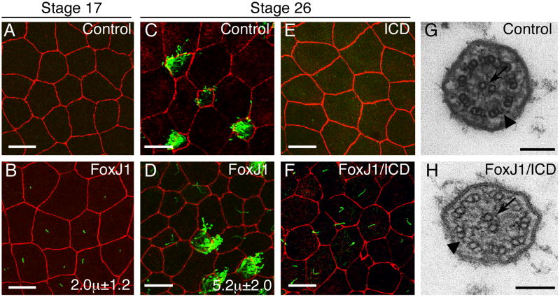
Figure 3.
XFoxJ1 RNA misexpression in surface epithelial cells induces ectopic cilia formation.
(A-F) Shown is a confocal image of the superficial epithelium in Xenopus embryos at the indicated stage, stained with antibodies to ZO-1 (Red) and acetylated-tubulin (green) to label cell borders and cilia, respectively. Embryos were injected at the two-cell stage with RFP RNA alone (A,C) with FoxJ1 and RFP RNA (B,D), with ICD and RFP RNA (E) or with FoxJ1, ICD and RFP RNA (F). Average cilia length in microns is indicated in B and D (n=15-20 cilia from 3 embryos). Scale bars are 20μm. (G,H) Transmission electron micrographs of a cilium in a multi-ciliate cell (G) or of an ectopic cilium (H) induced by FoxJ1 RNA in an ICD background (as in panel F). Arrows indicate the central pair and arrowheads indicate outer dynein arms. Scale bars are 100nm.

