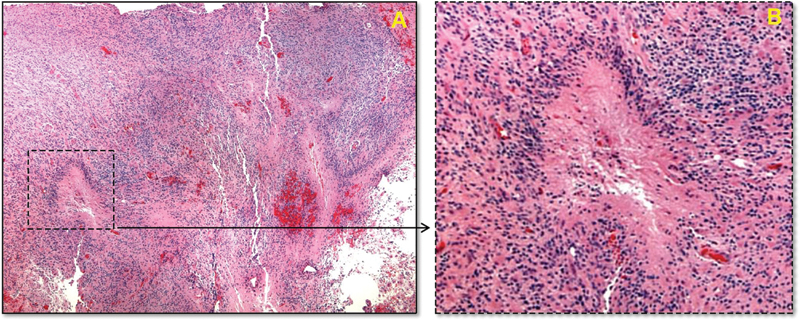
Fig. 2.

Histologic examination of the excised tissue. (A) Histologic section of the resected mass (hematoxylin and eosin stain) at low magnification, showing the serpentine pattern of necrosis and surrounding hypercellularity. (B) Higher magnification of the inset area showing a focus of necrosis, with the characteristic perinecrotic pseudo-palisading pattern that occurs with glioblastoma. Red discoloration occurs due to bleeding from necroses and microvascular proliferation.
