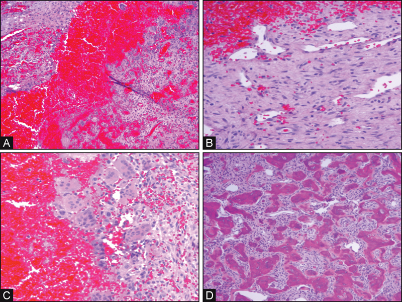
Fig. 4.

(A) Low-power view, variable size blood-filled spaces divided by fibrous septae. (B) Higher power view, loose fibrous tissue containing multiple capillaries and monotonous population of plump fibroblasts. (C) Small group of osteoclast-like giant cells. (D) Background of benign fibro-osseous proliferation (fibrous dysplasia versus ossifying fibroma).
