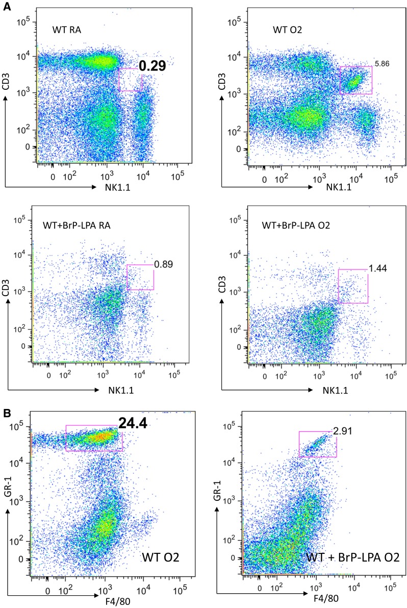
Fig. 5.

Flow cytometry analysis of pulmonary NKT cell and PMN populations after hyperoxia. a Lungs extracted from oxygen-exposed mice with and without BrP-LPA treatment were analyzed. Baseline iNKT cell populations in the lung did not differ between BrP-LPA treated and untreated wild type mice under normoxic conditions. After 72 h of 100 % exposure, animals show significant increases of iNKT cells compared to their baseline. After BrP-LPA treatment, animals exhibit only a minimal increase of pulmonary iNKT cells in response to hyperoxia. b Populations of GR-1+/F4/80- PMN-cells increase massively after oxygen exposure in lungs. When animals were injected with BrP-LPA prior to oxygen exposure PMN-cells only increased marginally
