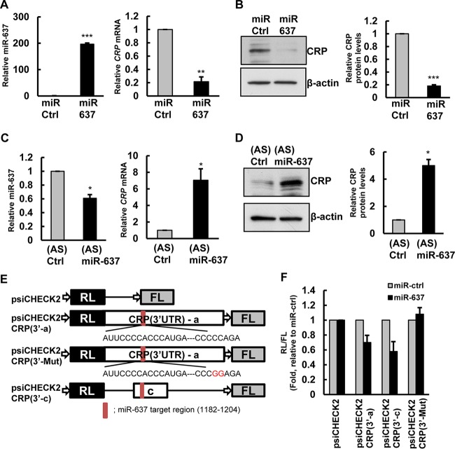
FIG 4.
miR-637 interacts with CRP mRNA. (A and B) Forty-eight hours after transfection with control miRNA (miR-Ctrl) or with miR-637, the levels of miR-637 (left) and CRP mRNA (right) were measured by RT-qPCR analysis (A), or protein levels were assessed by immunoblotting with anti-CRP and anti-β-actin antibodies, as a loading control (B). CRP protein levels were quantified from immunoblots and normalized to values for β-actin. (C and D) HeLa cells were transfected with antisense (AS) miR-637 or control miRNA. After 72 h, the levels of miR-637 (left) and CRP mRNA (right) were measured by RT-qPCR analysis (C), or the protein levels of CRP and control β-actin were analyzed by immunoblotting (D). CRP protein levels were quantified from immunoblots and normalized to values for β-actin. (E) Schematic of psiCHECK2-CRP dual-luciferase reporters. Red bar, predicted miR-637 binding site. (F) Twenty-four hours after transfection of HeLa cells with miR-637, cells were transfected with different CRP 3′-UTR luciferase reporter plasmids, and luciferase activity was measured (RL/FL ratio). The histograms represent the means and standard errors of the means from three independent experiments. *, P < 0.05; **, P < 0.01; ***, P < 0.001 (by Student's t test).

