FIG 2.
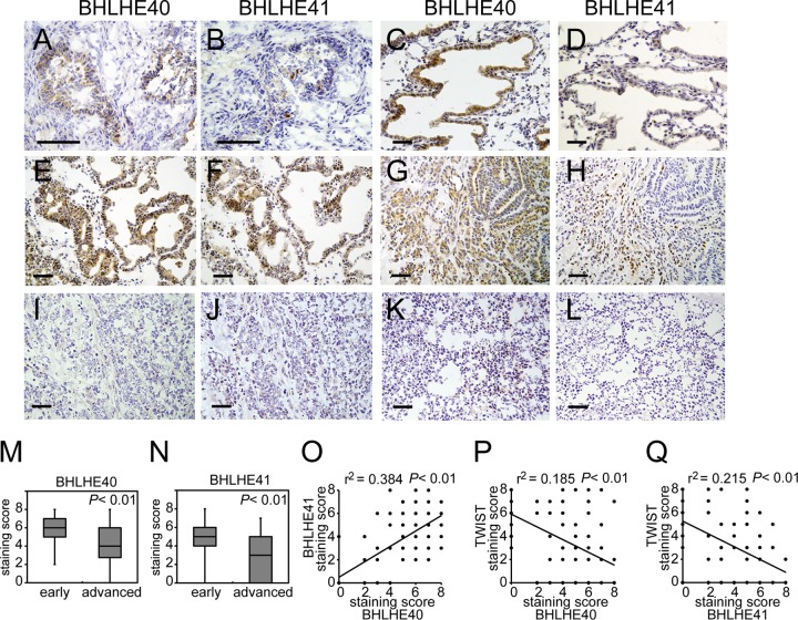
Immunohistochemistry of endometrial cancer specimens. Surgical samples from 86 HEC patients were analyzed for BHLHE40/41expression by immunohistochemistry. Representative results are shown. (A to D) Results are for the late proliferative phase (A and B) and the secretory phase (C and D) of normal endometrial tissue. (E and F) A grade 1 endometrioid adenocarcinoma (EAC) case at stage IA; (G and H) a grade 2 EAC case at stage IA; (I and J) a grade 3 EAC case at stage IB; (K and L) a grade 2 EAC case at stage IIIA. Immunohistochemical images with an anti-BHLHE40 antibody (A, C, E, G, I, K) and anti-BHLHE41 antibody (B, D, F, H, J, L) are shown. The scale bars represent 200 μm. (M and N) The staining scores of immunohistochemical images were analyzed. Eighty-six cases were divided into a group at the early stage (stage IA) and that at the advanced stage (more than stage IB). (O) The relationship between BHLHE40 and BHLHE41 staining levels from 86 specimens was analyzed by Pearson's product-moment correlation coefficient. r values are correlation coefficients. The significance of coefficients was determined by the F-test. (P and Q) The relationship between TWIST1 and BHLHE40 or TWIST1 and BHLHE41 staining levels was also analyzed by Pearson's product-moment correlation coefficient. A P value of <0.05 was considered significant.

