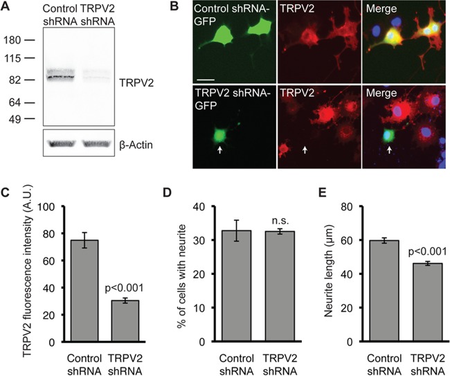
FIG 4.
Silencing of TRPV2 expression impairs NGF-induced neurite outgrowth. (A) Western blot analysis with the indicated antibodies of HEK293T cells transiently transfected with rat TRPV2 and either control or TRPV2 shRNA. (B) PC12 cells were transfected with control shRNA or TRPV2 shRNA. Twenty-four hours later, cells were treated with NGF (100 ng/ml) for 72 h. Cells were then fixed and immunostained for TRPV2 (red). GFP indicates a transfected cell (green), and nuclei were stained with Hoechst dye (blue). Bar, 50 μm. (C) Mean TRPV2 fluorescence intensity ± SEM for GFP-positive cells (control shRNA, n = 30; TRPV2 shRNA, n = 19) (P < 0.001 as determined by an unpaired t test). A.U., arbitrary units. (D) PC12 cells were transfected with control or TRPV2 shRNA, followed by 72 h of treatment with NGF (100 ng/ml). Fifty random images were obtained for GFP fluorescence and analyzed for the percentage of cells with neurites. Data represent the mean percentages of neurite-bearing, GFP-positive cells ± SEM from 3 independent experiments. n.s., not significant (P = 0.94) as determined by an unpaired t test. (E) Mean neurite lengths ± SEM for PC12 cells transfected with control shRNA (n = 362) or TRPV2 shRNA (n = 454) and treated with NGF for 72 h (P < 0.001 as determined by an unpaired t test).

