
FIG 1.
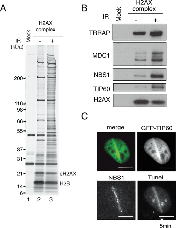
Identification of NBS1 in the H2AX complex. (A) The mock control and eH2AX complexes were immunoaffinity purified from the nuclear soluble fraction of HeLa cells that were not (−) or were (+) treated with 12 Gy IR, followed by a 5-min recovery. (B) Immunoblotting analyses were performed with anti-TRRAP, anti-MDC1, anti-NBS1, anti-TIP60, and anti-H2AX antibodies. (C) Accumulation of TIP60-GFP in GM02063 cells at sites containing DSBs after a 2-min recovery from laser UVA microirradiation. NBS1 staining was performed. Terminal deoxynucleotidyltransferase-mediated dUTP-biotin nick end labeling (TUNEL) staining was performed to detect DSBs induced by microirradiation. TIP60-GFP and TUNEL signals are shown in green and red, respectively, in the merged image. Scale bars, 10 μm.