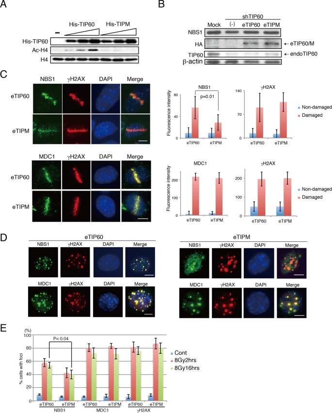
FIG 2.
Acetyltransferase activity of TIP60 is required for proper NBS1 localization to DNA damage sites. (A) His-TIP60 or His-TIPM was expressed in Escherichia coli, purified, and subjected to an in vitro H4 acetylation assay. (B) eTIP60 and eTIPM were expressed in MEFs stably expressing shTIP60. The expression of endogenous TIP60 (endoTIP60), reconstituted eTIP60 (Flag-HA-TIP60) and NBS1 was detected by anti-TIP60, anti-HA, and anti-NBS1 antibody, respectively. (C) Left, examples of microirradiated cells incubated for 4 h before fixation. Microirradiation experiment using eTIP60- or eTIPM-reconstituted MEFs was followed by immunohistochemistry analysis. Top, anti-NBS1 and anti-γ-H2AX antibodies; bottom, anti-MDC1 and anti-γ-H2AX antibodies. Right, the average fluorescence intensities from the undamaged regions or the damaged regions were quantified and plotted. The average intensities of NBS1 immunofluorescence were derived from 11 cells for TIP60-reconstituted cells and 10 cells for TIPM-reconstituted cells, and the average intensities for MDC1 immunofluorescence were derived from 8 cells for TIP60-reconstituted cells and 6 cells for TIPM-reconstituted cells. Examples of measured regions are shown in Fig. S2A in the supplemental material. See Materials and Methods for quantification details. (D) Immunohistochemistry analysis of MEFs reconstituted with TIP60 (left) or TIPM (right) following IR (16 h after 8 Gy). Indirect immunofluorescence analyses were performed using either anti-NBS1 and anti-γ-H2AX or anti-MDC1 and anti-γ-H2AX antibodies. (E) Percentages of cells that formed more than 10 foci of NBS1, MDC1, or γ-H2AX are shown. Cont, control; bar, 10 μm. Graphs show standard deviations.

