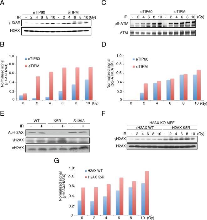
FIG 4.
Acetylation of H2AX K5 restricts γ-H2AX expansion. (A and B) γ-H2AX was monitored in eTIP60- or eTIPM-expressing MEFs (a), and the signals were quantified using ImageJ software (B). (C and D) Phospho-Ser1981 was monitored in eTIP60- or eTIPM-reconstituted MEFs (C) and quantified (D). (E) Ac-H2AX and γ-H2AX were monitored in cell lysates of H2AX KO MEFs reconstituted with H2AX WT, K5R, or S139A. Western blotting was performed with anti-Ac-H2AX(K5), anti-γ-H2AX, or anti-H2AX antibody. (F) H2AX KO MEFs were reconstituted with H2AX or H2AX K5R and subjected to Western blotting. Samples were prepared from cells irradiated with the indicated doses and incubated for 2 to 10 h. (G) Intensity ratios of the γ-H2AX to H2AX signals from the experiment whose results are shown in panel F.

