
FIG 6.
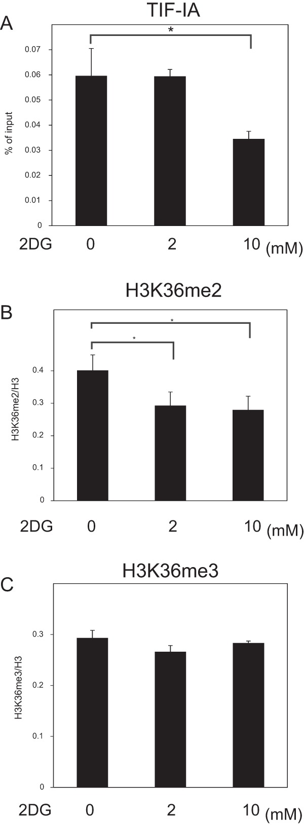
TIF-IA in rDNA promoter is decreased by 10 mM 2DG but not 2 mM 2DG. After MCF-7 cells were cultured 2 h in RPMI 1640 supplemented with 10% serum in the absence or presence of 2DG at the indicated concentrations, ChIP analyses were performed to detect TIF-IA (A), H3K36me2 (B), and H3K36me3 (C) in the rDNA promoter. The results of panel A were expressed as the percentage of input. The results of panels B and C are expressed as in Fig. 2B. All experiments were performed three times, and mean values with the standard deviations are indicated. *, P < 0.05.