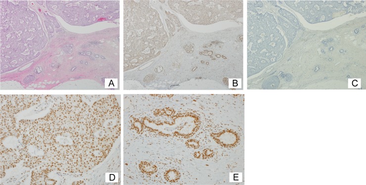
FIG 8.
Human breast cancer tissues express KDM2A. (A) Breast cancer tissue with triple-negative papillotubular carcinoma (hematoxylin and eosin staining; magnification, ×40). (B) Serial section of panel A stained with anti-KDM2A antibody, showing the expression of KDM2A in both neoplastic and nonneoplastic areas (×40). (C) Control serial section, in which the primary antibody was omitted (×40). (D) High magnification of the neoplastic area in panel B (×200). (E) High magnification of the nonneoplastic area in panel B (×200). Positive staining is brown, and counterstained nuclei are blue (B to E).

