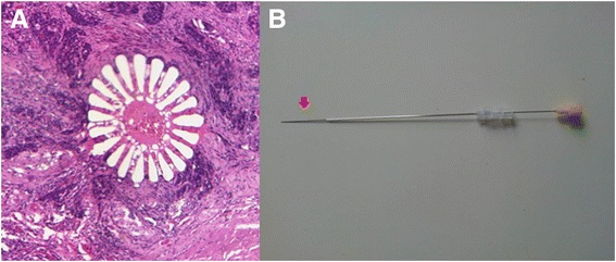
Fig. 2.

Sea urchin spine fiducial markers. a Example of a 5-μm-thick tissue section showing a sea urchin spine in the transverse plane in a haematoxylin and eosin (H&E)-stained histopathology slice at a spatial resolution of 1 μm/pixel. b 20-gauge spinal needle loaded with the sea urchin spine fiducial marker. The tip of the spine can be seen protruding from the needle (red arrow)
