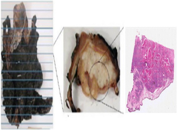
Fig. 4.

Slicing of excised specimen. A schematic diagram showing (left) the excised specimen, (centre) a ~5-mm-thick tissue slice and (right) a 5-μm-thick tissue slice that has been stained. A relevant section is taken from the 5-mm slice which is then embedded in paraffin prior to microtome cutting to obtain the 5-μm-thick tissue slices
